
How long should I wait?
Most elective revisions are planned after the previous nose has had enough time to reveal its stable shape—commonly around 12 months, and sometimes longer.
Read the timing section →
Revision rhinoplasty is not simply a second attempt at the same operation. Scar tissue, altered cartilage, previous incisions and changed airway support make diagnosis and planning more complex. The goal is to identify what can be improved, what must be protected and what limitations should be accepted.


Search this page’s medically reviewed answer library. All answers remain on this page and link to the full explanation.
Most elective revisions are planned after the previous nose has had enough time to reveal its stable shape—commonly around 12 months, and sometimes longer.
Read the timing section →Not automatically. The choice between remaining septal cartilage, ear cartilage and rib cartilage depends on how much structure and strength are required.
Compare graft sources →It may help when obstruction comes from residual septal deviation, nasal-valve narrowing, middle-vault collapse, scar bands or weak sidewalls.
Understand airway assessment →A small localised irregularity may need limited treatment; support loss, recurrent deviation, airway problems or several concerns usually need broader planning.
See the treatment scale →Scar cannot be removed without limit. It may need careful release, reshaping, camouflage or structural support while protecting the skin’s blood supply.
Read about scar and skin →Early recovery may look similar to primary rhinoplasty, but tip swelling and scar maturation can be slower and final refinement may take 12–18 months or longer.
Open the recovery timeline →This on-page guide cannot examine your nose, diagnose the cause of a problem, recommend surgery, calculate candidacy or replace consultation. Do not enter private medical details. Urgent pain, fever, skin colour change, heavy bleeding or sudden breathing deterioration needs direct medical care.
The earlier operation may have removed, shifted, scarred or weakened tissue. A responsible revision plan therefore starts by mapping what remains—not by promising a “perfect” second result.
Revision rhinoplasty—also called secondary rhinoplasty—is surgery performed after an earlier rhinoplasty, septorhinoplasty, septoplasty with external change, implant procedure or nasal reconstruction. The new concern may be something that never fully improved, something that returned during healing, or a new deformity caused by loss of support, scar contraction or airway narrowing.
Some patients are mainly concerned about a visible irregularity. Others have a combination of appearance and function: a pinched or collapsed middle vault, recurrent deviation, a drooping or over-rotated tip, nostril asymmetry, an over-reduced bridge, graft visibility, or difficulty breathing through one or both sides.
The first consultation should separate three issues: what is objectively present, what is still changing with healing, and what can realistically be improved without creating a new problem.
The visible complaint is only the starting point. Each pattern can have several structural causes, and more than one may coexist.
The nasal bones, septum, middle vault or tip may remain deviated, or scar and cartilage memory may pull the nose off-centre during healing.
Excessive dorsal removal or weakened septal support can produce a low bridge, open-roof irregularity, inverted-V shadow or unstable profile.
Lower-lateral cartilage damage, weak central support, scar contraction or excessive resection can distort tip shape and nostril support.
Thin skin, displaced cartilage, bone irregularity, scar tethering or implant/graft visibility may create localised surface problems.
Loss of rim support, scar tension or uneven tip/columellar change can alter the nostril margin and basal view.
Residual septal deviation, valve narrowing, middle-vault collapse, weak sidewalls, adhesions or untreated turbinate disease may contribute.
| Planning factor | Primary rhinoplasty | Revision rhinoplasty |
|---|---|---|
| Anatomical landmarks | Usually intact and easier to identify. | May be displaced, scarred, resected or hidden. |
| Cartilage supply | Septal cartilage is often available. | Septal cartilage may be limited after prior septoplasty or grafting. |
| Skin and scar | Unoperated skin envelope. | Scar thickness, tethering and altered blood supply may affect dissection and contour. |
| Airway | Baseline septum and valves are assessed. | Breathing may be affected by previous narrowing, collapse, adhesions or residual deviation. |
| Technique | Reshaping and preservation of native support. | May require restoration, replacement, camouflage and additional graft material. |
| Healing | Swelling pattern is variable but generally more predictable. | Tip swelling and scar remodelling may be more prolonged or uneven. |
| Goal setting | Change an unoperated nose. | Improve selected priorities within the limits of already altered tissue. |
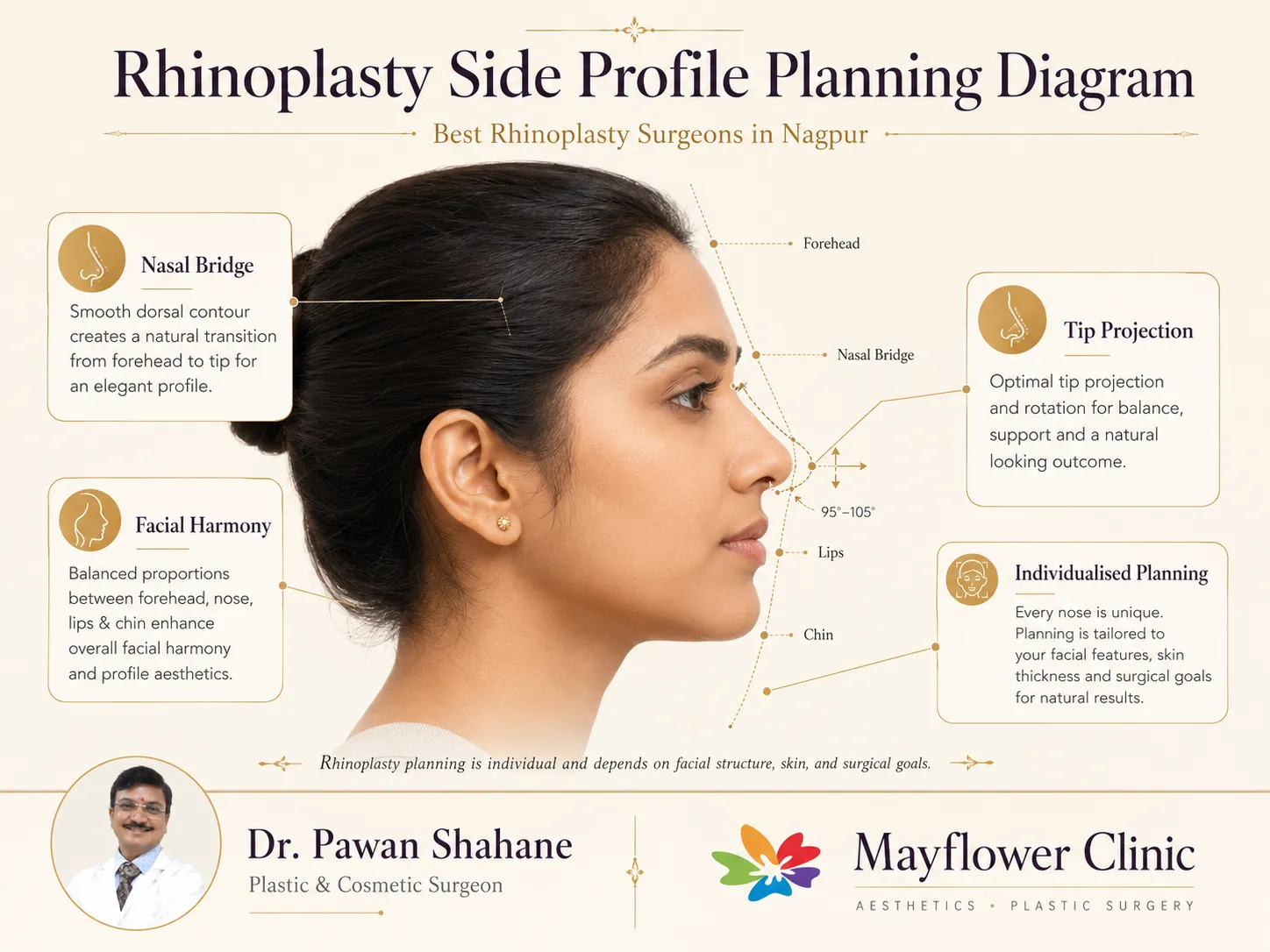
After rhinoplasty, swelling does not disappear evenly. The tip and supratip may stay firm or asymmetric for months, scar tissue continues to remodel, and the skin envelope gradually settles over the framework. An apparent early problem may improve, while a true structural problem may become clearer only after swelling reduces.
For most elective revisions, a common planning interval is about 12 months after the previous operation. Thick skin, multiple prior surgeries, extensive grafting or prolonged inflammation may justify waiting longer. This does not mean a patient must wait a year to seek advice; early consultation can document healing, check breathing and determine whether observation, steroid treatment, massage, medication or another non-operative measure is appropriate.
Bring preoperative photographs, operative notes, implant or graft details and a timeline of healing when available. Missing records do not prevent assessment, but known details can help.
List the one or two concerns that matter most. “Make it perfect” cannot guide safe surgery; a precise concern such as sidewall collapse or visible dorsal depression can.
Frontal, profile, oblique and basal views assess symmetry, bridge, tip, nostrils, columella, skin thickness and dynamic movement during smiling and breathing.
The septum, turbinates, internal and external valves, lining, scar bands, perforation and sidewall support are assessed. Endoscopy or imaging is used only when clinically indicated.
The surgeon estimates remaining septal cartilage, prior donor-site use, possible ear or rib graft need, implant status and skin-envelope limitations.
Computer imaging may help discuss direction, but it is not a guaranteed result. The plan should explain trade-offs, asymmetry limits and the possibility of further refinement.
| Pattern | Typical examples | Possible treatment direction | Important limitation |
|---|---|---|---|
| Observation / non-operative care | Early swelling, mild scar firmness, temporary asymmetry. | Time, photography, surgeon-guided care and treatment of inflammation when appropriate. | Cannot rebuild a true structural defect. |
| Limited touch-up | Small dorsal irregularity, localised scar tether or minor edge visibility. | Focused contour correction, scar release or camouflage in selected cases. | A “small” visible issue can still be difficult under thin skin. |
| Intermediate revision | Persistent tip asymmetry, alar retraction, residual hump or localised deviation. | Open or closed revision with selective grafting, suture work or osteotomy. | Healing can change adjacent areas, not only the targeted spot. |
| Major structural revision | Saddle deformity, pinched nose, valve collapse, recurrent severe deviation or failed implant/graft. | Framework reconstruction with multiple grafts, septal/valve work and possible ear or rib donor cartilage. | More swelling, donor-site considerations and residual asymmetry risk. |
Careful dissection may release tethering and expose altered structures. Fascia, perichondrium or finely prepared cartilage may be used for selected camouflage needs.
Depressions, over-reduction, implant problems, inverted-V deformity or irregular dorsal lines may require onlay grafts, spreader support, osteotomies or conservative smoothing.
Spreader grafts, spreader flaps, lateral-wall support or other techniques may be used when the middle third is narrow, unstable or functionally compromised.
Weak, pinched, drooping, over-rotated or asymmetric tips may need replacement of missing support, repositioning of cartilages, extension grafts or alar support.
Controlled bone cuts may be needed to correct a recurrent deviation, asymmetric bony vault, open roof or previously narrowed bridge. They are not used automatically.
Residual deviation, weak caudal support, perforation or lining deficiency can make revision more complex and may require a staged or specialised reconstructive plan.
The best graft source is not chosen by a slogan. It depends on what tissue remains, the amount and shape required, structural demands, donor-site trade-offs and the surgeon’s plan.
| Source | Common strengths | Common revision uses | Limitations / risks to discuss |
|---|---|---|---|
| Remaining septal cartilage | Straight, central donor material; no separate external donor site. | Spreader grafts, columellar or tip support, selected dorsal or caudal reconstruction. | May be insufficient, fragmented or already removed during earlier surgery. |
| Ear cartilage | Curved, elastic and useful for contour or alar support. | Alar batten/rim grafts, tip contour, selected camouflage and softer reconstruction. | Limited quantity and less suitable when long, rigid structural pieces are required; ear scar or contour change is possible. |
| Rib cartilage | Large volume and strong structural potential. | Major dorsal, septal, caudal or tip framework reconstruction when other cartilage is inadequate. | Chest scar, pain, contour change, rare pneumothorax, infection, warping, resorption or graft visibility/displacement. |
| Fascia / perichondrium / soft tissue | Thin biologic cover and camouflage. | Selected surface irregularities, thin skin or smoothing over cartilage. | Does not replace major structural support and can have variable thickness or resorption. |
Mayflower Clinic uses the term Max Lift / MX Lift rhinoplasty for a structural-support-oriented planning concept. In a primary nose, this may include coordinated tip support, projection, rotation, bridge contour, middle-vault stability and breathing assessment.
In revision rhinoplasty, the concept must be adapted to what remains. A patient with an over-rotated tip may need lowering and lengthening rather than more lift. A pinched nose may need widening and alar support. An over-reduced bridge may need augmentation. A drooping tip may need stronger central support. The name does not represent one fixed operation.
A straight-looking nose can still obstruct airflow, and a visibly imperfect nose may breathe well. Revision planning should identify the actual site or sites of resistance.
A persistent high, caudal or posterior deviation may contribute to obstruction. Remaining support must be protected while correcting the relevant segment.
Over-narrowing, hump reduction without adequate middle-vault support, scarring or upper-lateral cartilage collapse can reduce the valve area.
Pinched nostrils, alar retraction, weak lateral crura or dynamic sidewall collapse may require support rather than further reduction.
Enlarged turbinates, allergy, dryness, adhesions or lining damage may coexist. Not every breathing complaint is corrected by cartilage grafting alone.
Whistling, crusting, bleeding or obstruction can occur. Closure is complex, anatomy-dependent and not always indicated or successful.
Symptoms, side-specific obstruction, sleep and exercise impact should be documented. Aesthetic photographs alone cannot measure airflow improvement.
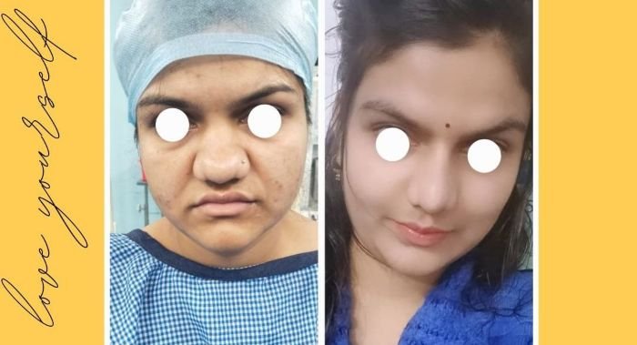
Previous surgery creates internal scar between the skin, cartilage and bone. In some areas this scar is thick and firm; in others the skin may be thin and tightly attached. Aggressive removal can injure the skin’s blood supply or create new irregularity.
Revision techniques may include careful scar release, conservative thinning in selected zones, repositioning of the skin envelope, structural grafting beneath a depression, and biologic camouflage with fascia or soft tissue. Thick skin may hide fine definition; very thin skin may reveal small graft edges.
Smoking, uncontrolled inflammation, repeated injections, previous infection, implant exposure and multiple prior operations can further affect healing. These factors must be disclosed before planning.
Bring old photographs, operative notes, implant information and a simple timeline. Write the two concerns that matter most, including any breathing symptoms.
The nose is examined from multiple views, palpated for support and evaluated during breathing. Skin, scar, valves, septum, turbinates and donor sites are reviewed.
Early swelling may need observation. A stable local concern may suit limited correction. Structural or functional problems may require full revision planning.
The surgeon discusses open or closed access, septal/ear/rib graft possibility, airway work, donor-site risks, hospital plan and what cannot be guaranteed.
Scarred anatomy is carefully exposed. The plan may be adjusted if the remaining framework differs from expectations. Stability and airway are reassessed during reconstruction.
Follow instructions for wound care, saline, medication, head elevation, activity restriction and any ear or chest donor site. Report red flags promptly.
Photographs and examination track contour, tip swelling and airway. Small asymmetries should not be judged too early; long-term review determines the stable result.
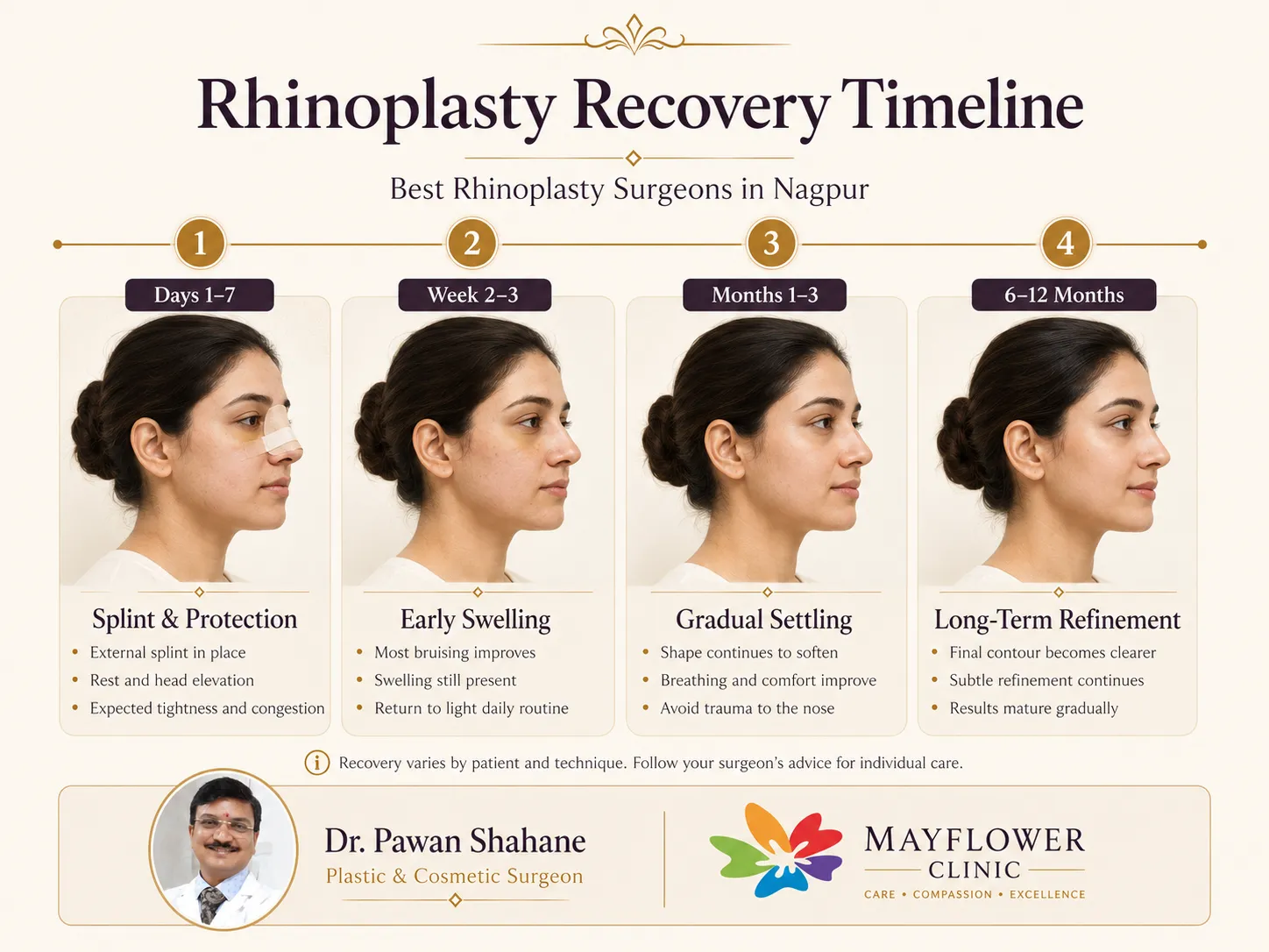
Revision rhinoplasty may involve previously operated skin, internal scar tissue, altered nasal support and sometimes ear or rib cartilage grafting. Follow the instructions given specifically for your operation, including wound and splint care, head elevation, saline or nasal care when advised, medication, activity restrictions and protection of any cartilage donor site.
Do not massage, press, manipulate or repeatedly compare the nose unless Dr. Pawan Shahane has specifically advised you to do so. Swelling can remain uneven for months, particularly around the tip, and early asymmetry does not necessarily represent the final result.
Contact the surgical team promptly for heavy or recurrent bleeding, rapidly increasing swelling, fever, pus, severe uncontrolled pain, grey/blue/black skin colour change, sudden major breathing difficulty, or unexpected problems at an ear or chest cartilage donor site.
Read the Mayflower Patient Care Guide →Bleeding, infection, anaesthesia complications, delayed healing, scarring, pain, numbness and reactions to medication can occur.
Residual asymmetry, visible edges, dents, bossae, over- or under-correction, recurrent deviation and dissatisfaction remain possible.
Nasal obstruction can persist or worsen; adhesions, septal perforation, valve narrowing, dryness or altered sensation may occur.
Warping, resorption, infection, displacement, visibility, extrusion or need for removal/revision can occur depending on material and tissue conditions.
Previously operated skin can be fragile. Prolonged swelling, skin thinning, vascular compromise, necrosis or poor scar quality are uncommon but important risks.
Ear contour change or scar, and chest pain, scar, contour irregularity, infection or rare pneumothorax may occur when donor cartilage is harvested.
These are educational and trust visuals—not claims of a specific revision outcome. Consented revision cases should be reviewed separately during consultation or added to the gallery only after verification.
“From the initial consultation to the post-operative care, Dr. Shahane demonstrated exceptional skill, professionalism, and a genuine care for my well-being.”Nishant Menghare
Google review · Rhinoplasty care
“He explains everything and how to take care. He support at every stage and do the surgery as planned.”Kunal Thakur
Google review · Rhinoplasty care
“I was always wanted to improve my nose but didn't know where to go. Then I got to know about Dr. Pawan Shahane.”Sushil Moon
Google review · Rhinoplasty care
These verified excerpts concern rhinoplasty care and are not presented as revision-specific patient testimonials or guarantees of outcome. Individual experiences and results vary.
Revision rhinoplasty is additional nasal surgery after a previous rhinoplasty, septorhinoplasty or other nose operation. It may address residual, recurrent or newly created concerns involving appearance, support or breathing.
For most elective revisions, surgeons prefer to wait until swelling and scar maturation are sufficiently stable—commonly around 12 months after the previous operation and sometimes longer. Earlier assessment is still useful, and urgent complications require prompt medical review.
Previous surgery may alter landmarks, remove or weaken cartilage, create scar tissue and change the nasal airway. The surgeon must work with the remaining framework and may need grafts or reconstructive techniques.
No. A small, well-localised irregularity may sometimes need limited correction, while support loss, recurrent deviation, airway collapse or multiple concerns may require a more extensive structural revision. Examination determines the scale of treatment.
It may improve breathing when obstruction is related to residual septal deviation, internal or external nasal-valve narrowing, middle-vault collapse, scar bands or weak sidewalls. Nasal allergy, sinus disease and turbinate problems may also need separate assessment.
Not everyone needs rib cartilage. Septal cartilage may be available in some patients, ear cartilage may suit curved or softer support needs, and rib cartilage may be considered when stronger or larger-volume reconstruction is required. Donor-site risks must be discussed.
Yes, when the amount, shape and strength are suitable. The graft source is chosen according to the defect, previous septal surgery, required support, skin thickness and the surgeon’s reconstruction plan.
No. Open access is often helpful in complex revisions because it exposes altered anatomy, but selected limited revisions may be performed through closed access. The approach should follow the required correction rather than a fixed preference.
Scar tissue cannot simply be removed without limit. Some scar is part of normal healing and may be closely attached to skin or cartilage. Revision surgery aims to release, reshape, camouflage or work around scar while protecting blood supply and skin quality.
Early splint, bruising and social recovery may resemble primary rhinoplasty, but swelling—especially at the tip—can be more persistent. Many patients plan about two to three weeks before important social events, while refinement may continue for 12 to 18 months or longer.
No. Revision surgery can improve selected concerns, but scar behaviour, skin thickness, graft healing, asymmetry and the limits of remaining tissue affect the result. A further touch-up can occasionally be required.
A septal perforation needs careful assessment of its size, location, symptoms, lining quality and remaining septal support. Not every perforation requires closure, and closure may need specialised reconstructive planning with no guarantee of success.
Fillers may camouflage selected depressions in carefully chosen patients, but they cannot rebuild weak support, correct major deviation or reliably improve breathing. Nasal filler also carries specific vascular risks and may complicate later surgical assessment.
There is no single revision-rhinoplasty price because complexity varies widely. Cost depends on the required reconstruction, graft source, anaesthesia, facility, functional work, investigations and follow-up plan. A written estimate should follow examination.
These publications support broad concepts about revision timing, altered anatomy, grafting, nasal obstruction, outcomes and complications. They do not replace individual clinical assessment.
Bring your previous photographs, operative details when available, and a clear list of appearance and breathing concerns. The consultation will focus on diagnosis, timing, remaining tissue, possible graft sources, risks and realistic priorities.
Medical Disclaimer: All surgical procedures carry inherent risks, and individual outcomes vary based on anatomy, previous surgery, scar tissue, skin type, graft availability and healing biology. Surgical results are not guaranteed. Formal in-person consultation with Dr. Pawan Shahane is required before any surgical decision. This page is for general educational purposes only and does not constitute medical advice or a treatment recommendation.

Case of the Month: Grade 3 Gynecomastia…

The Science of Facial Aging: Why Wrinkles,…