

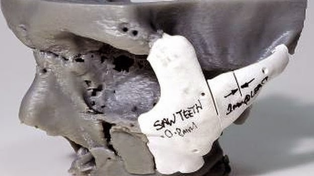
3D printing has moved from an experimental idea to a practical planning tool in selected areas of plastic and reconstructive surgery. Its main value is not that a printer “does the operation,” but that digital imaging and computer-aided design can be converted into physical models, surgical guides, prostheses or selected patient-matched devices that help a surgical team understand complex anatomy before treatment.
For plastic surgeons, this is most relevant when anatomy is difficult to visualize in two dimensions—for example in craniofacial trauma, jaw or orbital reconstruction, congenital deformity, complex bone defects and selected prosthetic applications. The technology can improve planning and communication, but it does not replace clinical examination, imaging interpretation, surgical judgment or multidisciplinary care.
Quick answer: what is 3D printing in surgery?
Medical 3D printing is a form of additive manufacturing. A digital three-dimensional file—often created from CT or other imaging data, or from computer-aided design—is converted into a physical object by building it layer by layer. Depending on the purpose, the final object may be:
- a patient-specific anatomical model for planning or teaching,
- a cutting, drilling or positioning guide,
- an external prosthesis,
- a template used to shape plates or grafts before surgery, or
- in selected regulated settings, a patient-matched implant or other medical device.
Where can 3D printing help in plastic and reconstructive surgery?
1. Craniofacial and maxillofacial planning
Complex facial fractures and defects can involve the orbit, cheekbone, upper jaw, lower jaw and several connected facial structures. A physical model can help the surgeon understand displacement, asymmetry and spatial relationships before entering the operating room. In selected cases, virtual surgical planning and patient-specific guides can also help translate the pre-operative plan to surgery.
Patients with facial-bone injuries can read more on our Maxillofacial Surgery in Nagpur page.
2. Reconstruction after trauma, tumour surgery or bone loss
When tissue or bone is missing, planning may involve restoring both function and contour. Three-dimensional models can help visualize the defect, compare the affected side with the unaffected side, estimate graft geometry and plan reconstruction. In some centres, patient-specific guides or implants are incorporated into the reconstructive workflow.
For a broader overview of this type of care, see Reconstructive Plastic Surgery.
3. Pre-contouring plates and planning grafts
In selected bony reconstructions, a model can allow plates or other fixation components to be planned or contoured before surgery. This may reduce intra-operative trial-and-error. Published work in mandibular reconstruction suggests that 3D-assisted planning can reduce operating and bone-flap ischaemia time in appropriate cases, although results depend on the procedure, planning system and surgical team.
4. External prostheses and patient-specific devices
3D printing is also used for external prostheses and for producing devices with complex geometry. Examples include selected cranial plates, orthopaedic components, prosthetic hands and patient-matched surgical guides. These applications require appropriate materials, manufacturing controls and device regulation; a simple 3D-printed planning model is not the same thing as an implant intended to remain inside the body.
5. Education and patient communication
A physical model can make complex anatomy easier to understand for trainees, surgeons and patients. It may help explain the location of a defect, the planned reconstruction and why more than one specialty may be involved.
What are the realistic advantages?
- Better spatial understanding: a complex three-dimensional deformity can be easier to appreciate as a physical model than on flat images alone.
- Procedure rehearsal and planning: the team can study the anatomy, test an approach and anticipate technical difficulties before surgery.
- Patient-specific planning: guides, prostheses and selected devices can be designed around an individual patient’s anatomy.
- Potential operating-room efficiency: pre-planning may reduce time spent shaping or trial-fitting components during selected procedures.
- Teaching and communication: models can help explain anatomy and reconstructive goals more clearly.
What are the limitations?
3D printing is a tool, not a guarantee of a better surgical result. Its usefulness depends on the clinical problem and on the quality of every step in the workflow.
- Imaging quality matters. A model is only as accurate as the imaging, segmentation and digital design used to create it.
- Not every operation needs it. Straightforward procedures may gain little from a printed model or guide.
- Cost and turnaround time vary. Complex design, manufacturing and sterilization may increase expense and preparation time.
- Materials have specific indications. A material suitable for an educational model may be completely unsuitable for surgical implantation.
- Regulation is important. Patient-matched implants and surgical guides are medical devices and must meet the relevant safety, quality and regulatory requirements.
- Digital planning does not replace surgical judgment. Tissue quality, blood supply, scarring, infection, nerve function and intra-operative findings cannot always be predicted from a model.
What about 3D bioprinting?
Bioprinting aims to place living cells and biomaterials in controlled three-dimensional patterns to create tissue-like structures. Research is progressing in areas such as cartilage, bone, skin, muscle and blood vessels. However, the ability to print fully functional replacement organs or complex living tissues for routine reconstructive surgery remains investigational. Challenges include vascularization, integration with the patient’s tissues, long-term viability, manufacturing standards and regulation.
Does Mayflower Clinic use 3D printing for every reconstructive case?
No. Most plastic and reconstructive decisions still begin with history, physical examination and appropriate imaging. In a complex case, the treating team may consider whether virtual planning, a patient-specific model, a guide or another digital workflow would add meaningful value. Availability and suitability depend on the diagnosis, hospital or manufacturing resources and the planned procedure.
For complex facial or reconstructive problems, the important first step is not requesting a particular technology—it is defining the anatomical and functional problem accurately and then choosing the most appropriate planning method.
Evidence and references
- U.S. FDA: Medical Applications of 3D Printing — overview of patient-matched models, guides, implants and prostheses.
- 3D-assisted surgery for reconstruction in the head and neck area — review of virtual planning and patient-specific models/guides in mandibular reconstruction.
- Sculpting the future: 3D printing in plastic surgery and prosthetic devices — review of current applications, benefits and limitations.
- Bioprinting and the future of reconstructive surgery — 2025 review of emerging tissue-engineering applications and remaining challenges.
Reviewed and updated: 17 September 2026. Educational content reviewed for Mayflower Clinic by Dr. Pawan Shahane, M.Ch. Plastic Surgery.
Medical disclaimer: This article is for general education. The role of 3D planning, printed models, surgical guides or patient-matched devices varies by diagnosis and facility. It does not constitute an individual treatment recommendation and does not guarantee surgical outcomes.


Comments are closed.